Day :
Session Introduction
Shruthi D K
Subbaiah Institute of Dental Sciences, India
Title: Bite Mark Analysis-A Important tool in the role of justice

Biography:
Dr. Shruthi D K has completed MDS in the year 2012 from CIDS, Rajiv Gandhi University, BDS from SDM College of dental sciences, Dharwad in the year 2008.Forensic Anthropology and odontology from Yenepoya University, Mangalore in the year 2018.Curently working as Associate Professor in the Department of Oral Pathology and Microbiology’. She has published 15 papers in reputed journals and PubMed. Active member of NGO Love without reason associated with helping children with cleft lip and palate. Involved in diagnosing and treating precancerous lesions and conditions in clinical practice.
Abstract:
Bite mark analysis is currently contentious. It is a vital area within the highly specialized field of forensic science and constitutes commonest form of dental evidence presented in criminal courts. The science of bite mark identification can be used to link a suspect to a crime. The present study puts forth comparing and analysis of photographs of bite marks using inter canine, inter molar, and perimeter measurements with the help of digitizer version 2.0 software. The study describes characteristics, mechanism of production, appearances of bite mark injuries, comparison techniques, technical aids used in the investigation of bite marks. It can be extremely useful for establishing link between victims and suspects or acquitting the innocents.
Clare Lowe
Newcastle Dental Hospital, Newcastle-Upon-Tyne, United Kingdom
Title: Adequate completion of radiology request forms at Newcastle Dental Hospital. (A two-cycle audit)
Biography:
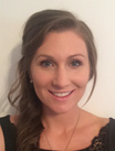
Clare Lowe is currently in the first year of her Dental Core Training at Newcastle Dental Hospital. She graduated from the University of Aberdeen in June 2016 with a Bachelor of Dental Surgery degree, and then went on to complete the diploma of Membership to the Faculty of Dental Surgery of the Royal College of Surgeons of Edinburgh in November 2017.
Abstract:
Objective: The usefulness of a radiological examination and its report can be reduced significantly if the clinical background and specific problem to be answered is not given in the request. Inadequate information can lead to mistakes in patient identification and delay in returning reports to the correct destination. The aim of this audit was to assess current request forms to determine if sufficient information was provided. The audit aims to ensure the quality of care provided to patients, and to identify ways to assist clinicians to provide adequate information when requesting a report.
Methods: Data was randomly collected from 53 patient records where a radiological request was made from the Oral Surgery department using the current request forms. Forms were analysed against eight criteria and recorded as either ‘criteria met’ or ‘criteria not me’. Data was recorded on a collection table and analysed to determine what percentage of radiology request forms could be deemed ‘adequate’, and when not, what were the failing criteria? A new form was then constructed considering the failings of the first cycle of data collection. The new radiology request form was then used for a period of 3 months and a second cycle of data collected.
Results: The first cycle of the audit revealed that 0% of request forms met the standard set, with 100% of forms omitting at least one of the criteria measured. Following the implementation of the redesigned form, the second cycle revealed that 70% of all forms met all criteria and could be deemed as adequate.
Conclusion: The new request form has dramatically improved the way the forms are completed. Marked improvement was noted in the information provided by clinicians on the new forms, showing that the new design helps to prompt clinicians to provide adequate information for reports to be generated.
Muy-Teck Teh
Institute of Dentistry, Queen Mary University of London, UK.
Title: From Transcriptome to a Pratical & Cost Effective Digital Cancer Test
Biography:
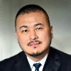
Dr Teh completed his PhD in 2000 at King’s College London followed by postdoctoral studies at Queen Mary University of London (QMUL). He is now a Senior Lecturer at the Centre of Oral Immunobiology and Regenerative Medicine, Barts and the London School of Medicine and Dentistry, QMUL. Dr Teh was awarded “Molecule of the Year 2010” for his pioneering research on FOXM1 in human cancer initiation. He has published over 50 papers in high impact journals and also as editorial board member of several reputed journals. His leads a clinical translational research group investigating disease biomarkers and mechanisms of molecular reprogramming.
Abstract:
Clinical translation of many cutting-edge technologies is hindered by their high costs and complexity despite highly promising results. Hence, affordability and practicality should be the core principles when developing a clinical tool. Big-data technological advances have produced large amount of biomolecular data for human cancers. However, the translation of these data into clinical use remains a challenge and impractical. In the attempt to resolve this problem, we have developed an affordable, practical and customisable cancer test - quantitative Malignancy Index Diagnostic System (qMIDS), which converts multiple gene expression levels (with SYBR-green based RT-qPCR) via an algorithm to provide a digital readout indicative of disease status. To date, we have collectively validated the qMIDS test on over 427 clinical head and neck samples from Europe and Asia, giving a diagnostic sensitivity/specificity/accuracy of ~90% compared to the gold-standard histopathology. We have further demonstrated the customisability and transferability of qMIDS test for diagnosing different squamous cancer types (vulva and skin). We also demonstrated the use of qMIDS for predicting the risk of drug resistance. We provided evidence that qMIDS was able to quantitatively and objectively stratify cancer aggressiveness/behaviour using a panel of biomarkers selected from transcriptomic databases. This study demonstrated an affordable method using a well-established and widely available qPCR technology for translating omics data into a practical and customisable clinical tool for disease diagnosis and prognosis.
Shruthi D K
Subbaiah Institute of Dental Sciences, Shimoga, Karnataka, India
Title: An early diagnosis of oral precancerous lesions and conditions, its role in oral cancer prevention

Biography:
Dr. Shruthi D K has completed MDS in the year 2012 from CIDS, Rajiv Gandhi Unieversity,BDS from SDM College of dental sciences, Dharwad in the year 2008.Forensic Anthropology and odontology from Yenepoya University, Mangalore in the year 2018.Curently working as Associate Professor in the Department of Oral Pathology and Microbiology’. She has published 15 papers in reputed journals and PubMed. Active member of NGO Love without reason associated with helping children with cleft lip and palate. Involved in diagnosing and treating precancerous lesions and conditions in clinical practice.
Abstract:
The study presents the result of local, systemic risk factors, peculiarities of clinical manifestation, quality of primary diagnosis of precancerous oral lesions. Oral cavity cancer accounts for approximately 3% of all malignancies. Most oral malignancies occur as squamous cell carcinoma. Many oral squamous cell carcinomas develop from premalignant conditions of oral cavity. To prevent malignant transformation of these precursor techniques have been developed to address this problem. The early detection of cancer is of critical importance because, survival rates markedly improve when the oral lesions are identified at an early stage. Hence the study emphasizes on the importance of early detection, diagnosis of precancerous lesions and its role in prevention of transforming onto oral cancers.
Wagner Breit
Maxillo Facial Surgeon in Brazilian Army
Title: Total Mandibular Reconstruction with Total Custom Titanium Prosthesis in Segmented Microvascular Fibula
Biography:
1.Markiewicz MR,Bell RB. Theuse of 3D imaging tools in facial plastic surgery. Facial Plast Surg Clin North Am 2011;19(4):655-682,ix
2. Haddock NT, Monaco C, Weimer KA, Hirsch DL,LevineJP,Saadeh PB. Increasing bony contact and overlap whith computer-designed offset cuts in free fibula mandible reconstruction. J. Craniofac Surg 2012;23(6):1592-1595
3.Essig H, Rana M, Kokemueller H, et al. Pre-operative planning workflow resulting in a pacient specifc reconstruction. Head Neck Oncol 2011;3:45
4.Wilde F, Plain M,Riese C,Schramm A, Winter K. Mandible recontruction whith patient-specifc pre-bent reconstruction plates: comparison of an in vitro study. Int J CARS 2012;7(1):57-63
5. Hidalgo DA. Titanium miniplate fixation in free flap mandible recostruction. 1989;23:498-507
6.Roser SM,Ramachandra S, Blair H, et al. The accuracy of virtual surgical planning in free fibula mandibular reconstruction: comparison of planned and final results. J Oral Maxillofac Surg 2010;68(11):2824-2832
7. Wild F ,Cornelius C-P,Schramann A. Computer-Assisted Mandibular Reconstruction using a Patiente-Specif Reconstruction Plate Fabricated with Computer-Aided Design and Manufacturing Techniques. Cranio Maxillo Fac Trauma Reconstruction 2014;7:158-166
Abstract:
The objective of this work is to show a new surgical protocol in high complexity reconstructions in the mandibular skeleton, predicting more stable results and a greater chance of rehabilitation with integrable bone implants.
This surgical case. is the first made in the world. Until then, we only have cases of the most varied types of mandibular reconstructions, either micro-vascularized, or autogenous grafts isolated or with plaques of reconstruction, not guaranteeing a stable long-term result for the patient mutilated by extensive segmentations caused by benign or malignant oral pathology .
Therefore, in this particular case, it shows a case of a patient with extensive ameloblastoma, who in a first surgical phase, was performed full mandibulectomy with a wide margin of safety and microvascular fibular graft with green-breasted fibular segmentation, grafted bone graft interposition bioss, rhBMP-2 and osteosynthesis with miniplates and 2.0 screws.
After a few months after surgery, the appearance of the microvascular fibular graft was morphologically in poor position, with vertical height exaggerated due to the high degree of angulation of the fibula, fibrosis in the stumps grafted with, bone instability to withstand chewing forces for future rehabilitation with osseointegrated implants.
Therefore, a new surgery, with extra-oral access, segmentation of the fibular graft in specific areas and studied in virtual planning and manufacture of the total mandibular prosthesis in custom titanium, with height and mandibular shape, thicknesses at strategic locations as a zone of traction and compression, as well as to predict the exact locations for placement of the implants and their complete rehabilitation.
Literature so far, there are no such extreme cases available. Only cases of mandibular hemi-prosthesis.
In this case, it is necessary to discuss the technique, virtual evaluation and imaging, in order to promote a rehabilitative surgery with extreme stability, predictability and establish normal functionality to the patient.
Salwa Regragui
Professor in orthodontics, Faculty of Dentistry, University Mohamed V in Rabat, Morocco
Title: Oral breathing: Study of prevalence and risk factors in a Moroccan population
Biography:
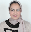
Salwa Regragui has completed her PhD and postdoctoral studies in Rabat Faculty of Dentistry in Moroccan Mohamed V University. She was the head’s of orthodontics department. She is the President of Moroccan association of orthodontics. She has published 6 papers in indexed journal:
“International Orthodontics”
1- Etude de la dysharmonie dento-dentaire à travers l’indice de Bolton dans une population Marocaine
2- Etude de la validité des tables de prédiction des dimensions des dents de Moyers, Tanaka et Johnston
3 – Etude des dimensions des Incisives latérales : responsabilité dans les dysharmonies dento-dentaires antérieures
4- Which method to measure dentomaxillary discrepancy?
6.Study of the adaptability of preformed orthodontic archwires to the average dental arch form of a Moroccan population
Abstract:
Introduction
Oral breathing is a dysfunction that sets in to compensate for deficient nasal ventilation thus favoring oral-cranial-facial malformations.
Due to the absence of epidemiological data on this dysfunction in Morocco, we conducted a study to determine its prevalence according to risk factors.
Materials and method
The study was conducted on schoolchildren in three regions with different climate and pollution levels; and was based on the use of a clinical diagnostic record.
Results and discussion
The prevalence of oral breathing, as well as the accompanying facial and dental changes, increase with the degree of humidity and pollution.
The comparison of our results with studies conducted in other countries with different climates, cultures and lifestyles has given rise to a very interesting discussion on this dysfunction, which deserves its integration into the health policy of countries
Isaac Firooze Moqadam
University Of Medical Sciences, Tehran
Title: Interleukin-10 gene polymorphisms in recurrent aphthous stomatitis
Biography:
Isaac Firooze Moqadam is a Board Certificated Periodontitis. He has several publications about oral pathology, oral lesions and periodontics.
Abstract:
Recurrent aphthous stomatitis (RAS) is a common oral inflammatory disease with unknown etiology in which the immune system seems to have a role in oral tolerance. Interleukin (IL)-10 is a cytokine synthesis inhibitory factor. Single nucleotide polymorphisms (SNPs) of IL10 gene could alter this cytokine production. The aim of this study was to investigate frequencies of IL10 alleles and genotypes in a group of individuals with RAS. Genomic DNA of 60 Iranian patients with RAS were typed for IL10 gene (C/A -1082, C/T -819, and C/A -592), using PCR-SSP method. Frequency of each allele and genotype was compared to control group. A significantly higher frequencies of the T allele at position -819 (p=0.006) and the A allele at position of -592 (p<0.001) were found in the patients with RAS group, when compared to the controls. IL10 GA genotype at position -1082 (p=0.007), CA genotype at position -592 (p=0.001), and CT genotype at position -819 (p=0.001) were significantly higher in the RAS patients. The results of this study suggest that certain SNPs of IL10 gene have association with predisposition of individuals to RAS. However, further multicenter studies should be conducted to confirm the results of this study.
Salwa regragui
Professor in orthodontics, Faculty of Dentistry, University Mohamed V in Rabat, Morocco
Title: STUDY OF variations of the DENTAL DIMENSIONS depending ON ANGLE OCCLUSAL CLASSES, SEX AND HOMOLOGOUS TOOTH
Biography:
Salwa Regragui has completed her PhD and postdoctoral studies in Rabat Faculty of Dentistry in Moroccan Mohamed V University. She was the head’s of orthodontics department. She is the President of Moroccan association of orthodontics. She has published 6 papers in indexed journal:
“International Orthodontics”
1- Etude de la dysharmonie dento-dentaire à travers l’indice de Bolton dans une population Marocaine
2- Etude de la validité des tables de prédiction des dimensions des dents de Moyers, Tanaka et Johnston
3 – Etude des dimensions des Incisives latérales : responsabilité dans les dysharmonies dento-dentaires antérieures
4- Which method to measure dentomaxillary discrepancy?
6.Study of the adaptability of preformed orthodontic archwires to the average dental arch form of a Moroccan population
Abstract:
The present study was performed on a sample of 140 Moroccan dental models in the orthodontics Department of the Faculty of Dental Medicine of Rabat. It consists of measuring of maxillary and mandibular dental-dimensional with digital caliper in samples which were separate into male and female teeth, left and right teeth of the same arch, occlusal classes I, II and III. Results are reported and statistically processed using SPSS version 18 software.
The aim of this study is to check the existence of a change in the dental dimensions according to the various parameters such as sex, right and left teeth or Angle occlusal classes.
The results are statistically significant only for maxillary arch in the Class I and III. (p<0,05)
However they are not significant between males and femals and confirme also a symmetry of the homolgous teeth (P> 0,05).
Farrukh Faraz
Maulana Azad Institute of Dental Sciences,India
Title: Bone Augmentation in Periodontics.
Biography:
Presently, Dr Farrukh Faraz is involved in teaching both the undergraduates & post graduate students at Maulana Azad Institute of Dental Sciences, New Delhi , India. Had completed his MDS in 2002 . Research expertise include involvement in an extensive research project under the aegis of Council of Scientific & Industrial Research, Govt. of India, where he is involved in Designing & manufacturing a state of art Implant system best suited to conditions of the Asian Subcontinent. Has more than 18 publications in National & International Journals.
Abstract:
Introduction: Regeneration has been the most sought after & also a rather elusive goal in Periodontics. This may be partly due to the mechanical, bacterial, thermal insults to which the oral cavity is subjected to time & again. Often the patient's report with soft and hard tissue defects result from a variety of causes, such as infection, trauma, and tooth loss. However, with the advances in biomaterials as well as techniques of Bone Allograft processing, there have been huge strides made in achieving the regenerative end result. With the advent of dental Implants, & to ensure prosthetically -driven dental implant therapy, reconstruction of the alveolar bone through a variety of regenerative surgical procedures has become more predictable. Reconstructive osseous surgeries include socket preservation, sinus augmentation, and horizontal and vertical ridge augmentation. The scope of use bone grafts in Periodontics is ever increasing Therefore, this case series aims at illustrating the application of different Bone grafts & their substitutes in varied situations for achieving optimal results for various therapies.
Conclusion: There exist many techniques & graft materials suited for different situations & achieving different objectives. An evidence based approach & extensive literature review could help the clinician in judicious treatment planning to achieve the desired objective.
Faisal Al Rumaihi
Prince Sultan Military Medical City, Saudi Arab
Title: Case Report of Impacted Bilateral Mandibular Fourth Molar
Biography:
Al Rumaihi Faisal has completed his BDS in 1988 in King Saud University in Riyadh KSA and Residency program at RKH Hospital 1989 advance certificate in restorative and cosmetic dentistry in 1995 at Boston University, USA and Ph.D degree at Boston University Goldman School of Graduate Dentistry USA. He is consultant restorative in dentistry in Prince Sultan Military Medical City in Riyadh. He is director of Restorative section in dental clinic and have a years of teaching and clinical supervision experience.
Abstract:
Supernumerary teeth is a rare dental anomaly in maxilla and mandible can be classified by shape and by position in the jaw. It might cause complication such caries, perio dental disease, and delay or impaction of permanent teeth.
Supernumerary tooth or hyper dontia is not as common as hypodontia. The prevalence in primary dentition .2 to .8 % and in the permanent dentition .5 to 5.3% with geographic variation.
The fourth molar is a kind of super numerary tooth they have been classified as a type of paramolar or disto molars tooth.
This case report of a 20 years old female pt. (Medically fit) come to the dental clinic complaining from pain in the lower right quadrant upon clinical examination and routine radiographic examination revealed impacted third molar and un erupted bilateral mandibular fourth molar orthopantogram (OPG) xray should this rare case of un erupted bilateral disto molar in mandible without any associated syndrome.
This case report discuss the diagnosis and treatment of this rare case of impacted bilateral mandibular disto molar and in what condition shall we keep the fourth molar or extracted.
Mohamed Hamdy Helal
Oral Diagnosis & Oral Radiology, Faculty of Dentistry, Tanta University, Egypt.

Title: Evaluation of Osseous Response after Placement of Experimental Implants in Femur of Diabetic Rabbits Using Piezoelectric Osteotomy.
Biography:
Mohamed hamdy has completed his B.D.S at the age of 22 years from Tanta University and now about finishing M.D in oral medicine , periodontology , diagnosis, and oral radiology.
Abstract:
Objective: To evaluate bone response at the interface bone – implant using piezoelectric osteotomy in diabetic rabbit’s femur, and com- pare with results using conventional osteotomy technique.
Material and Methods: Twenty New Zealand mature male rabbits with an average weight of (2-3.5 kg) were induced for diabetes using Streptozocin (STZ) drug, then diabetic rabbits had been controlled using insulin injection and were divided into 2 groups; The 1st group of 10 rabbits received implants prepared by conventional drills while the 2nd group 10 rabbits received implants prepared by Piezo sur- gery(PS). All rabbits were euthanized one month after the surgery. Samples were evaluated histologically using stereomicroscope, light microscope, then histomorphometrical analysis was done in order to estimate bone to implant contact (BIC).
Results: The histological results of our study using stereomicroscope and light microscope in group (A): implants showed Osseo integration with direct bone-implant contact visible at most parts of the cortical bone after 4 weeks’ period of follow up. However, small areas showed minimal gap spaces along the implant bone interface. With no observed signs of an in ammatory or foreign body reaction. While in group (B): Implants showed nearly complete Osseo integration with direct bone-implant contact visible at the cortical bone. Moreover, there was obvious thick layer of newly formed lamellar bone around the implants. Our histomorphometric analysis results of BIC was measured on the digitalized histological photomicrograph and expressed in pixels. PS showed non-signi cant increase in the percentage of the mean BIC at (p=0.268) compared to conventional technique.
Conclusions: PS permitted new bone formation for Osseo integration of titanium implants in controlled diabetics rabbits and a orded results similar to those of conventional technique. PS can be considered a feasible alternative in dental implant in diabetics.
Tarek Kasem
Salma Albati Minstry of Health, Saudi Arbia
Title: Platele-Rich Fibrin in Treatment of Central Giant Cell Granuloma
Biography:
BDS1995…………….MDS oral maxillofacial 2000.
Working in Saudi Arabia since 2001.
Damascuss university
Working Expernices:
- 2001- 2006 Chief of Dental Department in Alrass Genral Hospital , Ministry of Health, Saudi Arbia.
- 2001-2018 Oral maxillofacial specialist in Al Rass Genral Hospital.
- Coverning On-Call Of Emergency Room and Clinic All The Months Since 9 Years.
- Member in International Association Of Oral Pathology.
- Member in International Association Of Dental Truma.
- Attended 10 asian conferences of oral maxillofacial surgery in 2010 and a course of implant in Malysia noble biocare.
- Shared orthogantic surgery course and Course auto-tooth garft in south korea 2012
- A course in bern university 2015.
- Tow case report articles are puplished in international journals of scinetific and engeering research.
Abstract:
Background:
Platelet rich fibrin is a natural alternitave used to reduce inflamation and improve the healing process. It has recently been used in dentistry for that pupose which show low risks and favorable results.
Material and method:
A patient aged 26 years old, had central gaint cell granuloma in the left mandiblular angle. biopsy was collected before surgery to conferm the diagnosis. Flap was retracted and Lesion site was filled with patient’s platelet that was previously taken from the patient's blood. The sugical site sutured and the patient instructed. The patient was followed up after a week, and 3 months.
Result:
The result showed good bone and soft tissue healing with no opertive or post-operative comliactions. After three months, the bone regeneration was great with no recurrence.The follow up plane is still in progress.
Conclusion:
The Platelet rich firbrin can be used as an effective and satisfactory mean in treatment of some oral pathologies among them central giant cell granuloma.
Faraneh Abdolhoseinpour
Pedodontist ,Mashhad University Of Medical Sciences
Title: Evaluation of antioxidant efficacy of Purslane extract in Patients with Recurrent Aphthous Stomatitis: a randomized, placebo-controlled, triple-blinded, clinical
Biography:
Faraneh Abdolhoseinpour has got her DDS at 2012.She has finished her pediatric dentistry specialty at 2017.Now she is a Board Certificated Pedodontist at private practice.she has several publications about cleft lip and palate , primary dentition and also oral pathology. Recently (march 2017) she had a oral presentation in Rome (Italy).
Abstract:
This herbal medicine is considered a rich source of antioxidants with anti-inflammatory effects. The purpose of this study was to evaluate the effectiveness of purslane in treatment of recurrent aphthous stomatitis (RAS) and also it ̓s effect on antioxidant level. Materials and methods: 50 patients were selected for this randomized triple-blind placebo-controlled trial. All subjects were randomly divided in to two groups, one group received purslane (n=25) and another group, placebo (n=25) for 3 month. Superoxide dismutase (SOD), glutathione peroxidase (GSHPx) and total antioxidant status (TAS) was measured in plasma at baseline and after 3 month of treatment. Also pain intensity based on the visual analogue scale (VAS), the mean interval between lesion, number of lesions and the mean duration of complete healing at baseline and in month 1, 2 and 3 were recorded. Statistical analysis was performed by using Mann-Whitney and T-test. Results: A significant decrease in pain intensity in VAS scores was seen after treatment in intervention group (p<0.001). The mean duration of complete healing showed significant differences (P<0.001) between the two groups. The mean interval between lesions also showed significant differences (P<0.001) among the intervention group (33.12 days) compared with the placebo group (17.88 days). No significant differences were found regarding the number of lesions, level of erythrocyte GSHPx, TAS and SOD. No serious side-effects occurred in either of groups. Conclusions: According to this study, purslane is clinically effective in treatment of RAS (number of lesions, pain intensity and duration of healing) although it is unable to change the level of antioxidants.
Nada Al Hussain
King Khalid University, Iran
Title: A descriptive study of oral findings among psoriasis patients
Biography:
Nada has completed her BDS at the age of 24 years from King Khalid University. She is a demonstrator in oral and Maxillofacial surgery at king Khalid University. Previously, Nada worked as a volunteer in prince sultan military hospital, department of maxillofacial for one year.
A Member of Saudi Society of Oral and Maxillofacial surgery and representor of Society in south regain of Saudi Arabia.
Abstract:
Psoriasis is a chronic, immune-mediated inflammatory skin disease. Psoriasis is estimated to affect about 2–4% of the population in most western. The commonest type is psoriasis vulgaris, in which there are well-delineated pappulosquamous plaques. Aim of the study was to find out the distribution of oral lesions among psoriasis patients. Our Methodology was to undertake a cross sectional study to understand the distribution of oral lesions among psoriasis patients. The study also dealt with other factors such as blood group, co morbidities, immunoglobulins and demographic characteristics. The results were tabulated using SPSS software.Our study has lent support to the theory that psoriatic patients in general are presented with more oral lesions than non-psoriatic subjects (21). According to our study Geographic tongue was seen in 62.38% of the population, Fissured tongue in 3.96% of the population. Both fissured and Geographic tongue together was present in 23.76% of the population. Pigmentation was seen in 5.94% of the population. Considering the possibility of the presence of a shared genetic basis for psoriasis and GT is one of the possible explanations for the frequent occurrence of GT in psoriatic patients.
Josh Delacruz
University of Winnipeg,Manitoba, Canada
Title: Preventing Oral Cavity Cancers by Reducing Radioactive Particle Delivery in Cigarettes Using a Novel Filter Device

Biography:
Josh Delacruz is enrolled in a pre-dentistry program leading to a bachelor of science degree from the University of Winnipeg in Canada. He grew up in a rural area of Canada with high tobacco use and saw first hand the negative effects tobacco had with friends and loved ones. He has presented his data at different research gatherings at the provincial and national levels where he received medals and scholarships for his grassroots efforts on this little known topic.
Abstract:
In 1998 major American tobacco corporations were sued and became legally obligated to disclose their internal documents as part of the court-ordered Master Settlement Agreement. These documents revealed that as early as the 1960’s, the tobacco industry was aware that phosphate fertilizer was radioactively contaminated with Polonium 210. However, these tobacco companies continued to use this type of fertilizer, knowingly selling contaminated tobacco products to consumers. Radioactive particles from smoke inhalation can become lodged in the tissues of the oral cavity and can damage DNA structure, leading to cancer cell development. After conducting a randomized, controlled and blinded screening experiment on popular cigarette brands, it was found that all brands tested had alarming levels of radioactivity, exceeding internationally recognized thresholds for surface contamination. The most radioactive brand was ‘Winston’, the least being ‘Camels’. Using a Welch’s t-test, all brands demonstrated significantly higher radioactivity than blank placebos (p<0.05). A novel cigarette pre-filter was designed and tested on the most radioactive brand of cigarettes and was found to be an excellent filter against radioactive particles. Radioactive particle delivery was reduced by a statistically significant amount (p<0.003) using this novel, cost effective and simple zeolite pre-filter . According to the U.S. Surgeon General, radioactive contamination is responsible for 90% of all tobacco related oral cancers. Very little research effort is being done currently, although this study may encourage others to revisit the inconvenient truth about cigarettes and its negative impact in the field of dental health.
